Arteries Diagram ~ Coronary Arteries And Heart Disease. Human body artery diagram in detail. The neck is supplied by arteries other than the carotids. Each of these arteries delivers blood to the leg and continues into the foot, with the posterior tibial and fibular arteries forming the plantar arteries and plantar arch that supply blood to the bottom of the foot and toes. The large arteries leaving the heart and then makes around the body is provided by the beating heart and by blood pressure. This is known as the main pulmonary artery or pulmonary trunk.
This is a list of arteries of the human body. Blood flow through the right side of the heart involving the following cardiac structures: 5 out of 5 stars. Learn vocabulary, terms, and more with flashcards, games, and other study tools. Digital arteries arterial blood flow chart artery lateral femoral circumflex.

Peripheral nervous system 23p image quiz.
After receiving blood directly from the left ventricle of the heart, the. 5 out of 5 stars. An artery is an elastic blood vessel that transports blood away from the heart. These arteries and their branches supply all parts of the heart muscle with blood. The right and left subclavian arteries give rise to the thyrocervical trunk. The main pulmonary artery splits into the right and left pulmonary arteries, which we will better see in later images. Arteries and veins are two of the body's main type of blood vessels. The coronary arteries wrap around the outside of the heart. Size, location, and orientation the modest size and weight of the heart give few hints of its incredible strength. Superior vena cava (svc), inferior vena cava (ivc), right atrium (ra), tricuspid valve (tv), right ventricle (rv. Next, we have the blood vessel responsible for carrying deoxygenated blood from the right side of the heart (right ventricle) to the lungs. Arteries of the head and neck diagram art print vintage anatomy art print on tea stained paper dog art dog s wfh office art. Arteries are the blood vessels that carry blood away from the heart, where it branches into even smaller vessels.
Only 3 available and it's in 2 people's carts. The cardiovascular system consists of the heart, blood vessels, and the approximately 5 liters of blood that the blood vessels transport. Each of these arteries delivers blood to the leg and continues into the foot, with the posterior tibial and fibular arteries forming the plantar arteries and plantar arch that supply blood to the bottom of the foot and toes. Finally, the smallest arteries, called arterioles are further branched into small capillaries, where the exchange of all the nutrients, gases and other waste molecules are carried out. Over the years, cholesterol plaques can narrow the arteries supplying blood to the heart.

Heart posterior 20p image quiz.
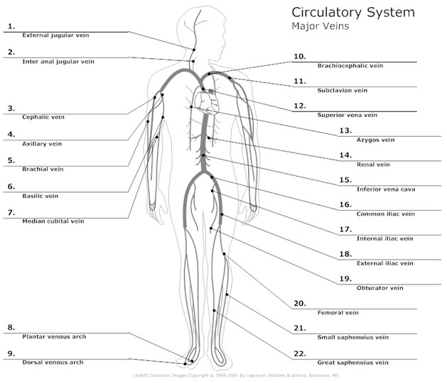
An artery is an elastic blood vessel that transports blood away from the heart. In this image, you will find right gastric artery, common hepatic artery, celiac trunk, left gastric artery, splenic artery, splenic vein, pancreas, suprarenal vein, renal vein, renal artery, inferior mesenteric vein , gonadal vein, gonadal artery, two alternative position of artery, left colic artery. 5 out of 5 stars. Veins are the blood vessels present throughout the body. Head/neck arteries 13p image quiz. Coronary arteries supply blood to the heart muscle. These vessels are channels that distribute blood to the body. The arteries' smaller branches are called arterioles and capillaries. Digital arteries arterial blood flow chart artery lateral femoral circumflex. Human body artery diagram in detail. The cardiovascular system consists of the heart, blood vessels, and the approximately 5 liters of blood that the blood vessels transport. This is known as the main pulmonary artery or pulmonary trunk. The triangles of the neck.
Size, location, and orientation the modest size and weight of the heart give few hints of its incredible strength. 5 out of 5 stars. These vessels are channels that distribute blood to the body. Only 3 available and it's in 2 people's carts. Each artery is a muscular tube lined by smooth tissue and has three layers:

Next, we have the blood vessel responsible for carrying deoxygenated blood from the right side of the heart (right ventricle) to the lungs.
The anterior tibial artery forms the arcuate artery and its many branches to supply blood to the top of the foot. Finally, the smallest arteries, called arterioles are further branched into small capillaries, where the exchange of all the nutrients, gases and other waste molecules are carried out. Arteries are components of the cardiovascular system. pulmonary artery sling can be treated surgically. In this image, you will find external carotid artery, internal carotid artery, vertebral artery, aorta and arch, pulmonary artery, cardiac artery, thoracic aorta, celiac trunk, superior mesenteric artery, renal artery, gonadal artery, inferior mesenteric artery, common iliac artery, external iliac artery. Size, location, and orientation the modest size and weight of the heart give few hints of its incredible strength. Veins are the blood vessels present throughout the body. 5 out of 5 stars. Smartdraw includes 1000s of professional healthcare and anatomy chart templates that you can modify and make your own. Superior vena cava (svc), inferior vena cava (ivc), right atrium (ra), tricuspid valve (tv), right ventricle (rv. The large arteries leaving the heart and then makes around the body is provided by the beating heart and by blood pressure. This is the opposite function of veins, which transport blood to the heart. Learn vocabulary, terms, and more with flashcards, games, and other study tools.
